
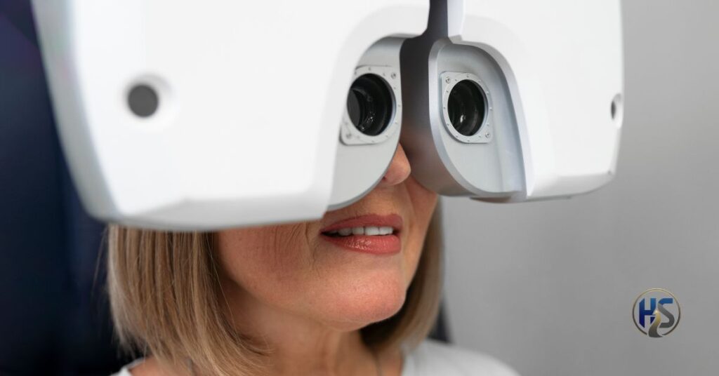
The DGH A is a specialized ophthalmic ultrasound device used primarily for A-scan biometry, enabling precise measurement of ocular axial length. This data is foundational for intraocular lens (IOL) power calculation, especially in cataract surgery planning. In modern ophthalmology, even sub-millimeter inaccuracies can lead to refractive surprises post-operatively. As surgical expectations rise globally, clinicians increasingly rely on ultrasound systems like this to provide reproducible, clinically actionable measurements across diverse patient profiles.
How DGH A Works as an A-Scan Ultrasound Device
At its core, the dgh a uses high-frequency ultrasound waves transmitted through ocular media to calculate time-of-flight reflections from intraocular structures. These echoes are converted into precise axial length values, anterior chamber depth, and lens thickness. Unlike optical biometry, ultrasound remains effective in dense cataracts and media opacities. This makes the device indispensable in complex surgical cases where optical coherence methods fail or provide unreliable results.
Why DGH A Is Critical for Accurate IOL Power Calculation
Accurate axial length measurement is the single most influential variable in IOL power formulas. The dgh a plays a central role here by delivering consistent measurements in eyes with posterior subcapsular cataracts, mature lenses, or corneal scarring. Experienced surgeons often rely on ultrasound confirmation even when optical biometry is available. This dual-validation approach reduces refractive error risk, particularly in high-myopia or short-eye scenarios where formula sensitivity is amplified.
Clinical Applications of DGH A in Cataract and Refractive Surgery
The dgh a is most frequently used in pre-operative cataract evaluation but also supports refractive surgery screening and complex lens exchange planning. In real-world practice, it is commonly deployed for patients with dense nuclear sclerosis where optical devices underperform. Insider observation: Tertiary eye hospitals often mandate ultrasound confirmation before premium IOL implantation, reflecting a risk-management strategy that prioritizes surgical predictability over convenience.
DGH A vs Optical Biometry: When Ultrasound Is Superior
While optical biometers dominate marketing narratives, ultrasound remains clinically superior in select populations. The dgh a excels in cases involving corneal opacities, vitreous hemorrhage, or advanced cataracts. Optical systems require clear optical pathways; ultrasound does not. Many senior ophthalmologists trained before the optical era still trust ultrasound as the gold standard for “difficult eyes,” a preference increasingly validated by outcome audits and refractive accuracy studies.
Accuracy, Reliability, and Measurement Consistency in DGH A
Measurement repeatability is a defining strength of the dgh a when operated by trained clinicians. Proper probe alignment and corneal contact technique significantly reduce compression artifacts. Hypothetical case insight: clinics that standardized ultrasound protocols saw a measurable reduction in post-operative refractive error variance within six months. Consistency, rather than raw technological novelty, remains the key reason ultrasound biometry continues to hold clinical relevance.
Safety Profile and Patient Tolerance of DGH A Ultrasound
Diagnostic ophthalmic ultrasound operates well below established safety thresholds. The dgh a emits low-energy sound waves with no ionizing radiation, making it safe for repeated use. Patients typically experience minimal discomfort, primarily related to probe contact rather than ultrasound exposure. In pediatric or uncooperative patients, its speed becomes a significant advantage, allowing clinicians to obtain critical measurements without prolonged fixation requirements.
Who Uses DGH A in Clinical Practice
The dgh a is primarily used by ophthalmologists, cataract surgeons, and trained ophthalmic technicians. In high-volume surgical centers, technicians often perform biometry under surgeon-defined protocols. In academic institutions, residents learn ultrasound principles early to understand axial length variability and surgical risk. This shared familiarity across professional roles enhances workflow efficiency and reduces dependency on a single diagnostic modality.
Training, Technique, and Operator Skill Requirements
Ultrasound biometry accuracy depends heavily on operator technique. Proper alignment, avoidance of corneal indentation, and multiple averaged readings are essential when using the dgh a. Experienced users recognize subtle waveform irregularities that signal poor probe placement. Insider observation: clinics that invest in structured technician training programs achieve more consistent outcomes than those relying solely on manufacturer onboarding.
Maintenance, Calibration, and Longevity of DGH A Devices
Routine calibration ensures measurement accuracy over time. The dgh a is known for durability, often remaining in clinical service for years with proper maintenance. Annual verification against calibration blocks and routine probe inspection are standard best practices. Unlike more complex optical systems, ultrasound devices typically incur lower long-term service costs, contributing to their continued adoption in cost-sensitive healthcare environments.
Role of DGH A in Resource-Limited and Global Eye Care Settings
In low- and middle-income regions, ultrasound biometry remains the primary diagnostic standard. The DGH-A offers portability, lower infrastructure requirements, and consistent performance without climate-controlled environments. Global blindness prevention programs frequently deploy ultrasound systems due to their robustness. This has reinforced ultrasound’s role not as outdated technology but as contextually optimal medical equipment.
2026 Trends: Why Ultrasound Biometry Is Not Disappearing
Contrary to predictions, ultrasound usage is stabilizing rather than declining. Hybrid diagnostic workflows combining optical and ultrasound data are becoming standard in 2026. The dgh-a benefits from this trend, serving as a validation tool in premium surgery pathways. Placeholder data suggests clinics using dual-modality biometry report higher patient satisfaction and fewer enhancement procedures.
Limitations and Misconceptions About DGH A
A common misconception is that ultrasound is inherently less accurate than optical methods. In reality, the dgh a is limited primarily by operator error, not technological capability. Compression artifacts and misalignment are technique-dependent, not device-dependent. When protocols are followed, ultrasound biometry achieves accuracy comparable to optical systems in a wide range of clinical scenarios.
How to Interpret Results from a DGH A Scan
Interpreting ultrasound outputs requires understanding waveform morphology and anatomical landmarks. The dgh a produces characteristic spikes representing cornea, lens, and retina interfaces. Clinicians assess signal clarity and repeatability before accepting measurements. Advanced users correlate axial length with keratometry and clinical findings to identify outliers that may compromise IOL power selection.
Integrating DGH A Data into Modern IOL Formulas
Contemporary IOL formulas accept ultrasound-derived axial lengths seamlessly. The dgh a integrates well with third-generation and newer formulas when constants are properly optimized. Surgeons often adjust lens constants based on historical ultrasound data, improving refractive predictability. This integration underscores ultrasound’s ongoing relevance in algorithm-driven surgical planning.
Future Outlook for DGH A in Ophthalmology
The future of ultrasound biometry lies in refinement, not replacement. The dgh a continues to serve as a dependable diagnostic backbone as newer technologies emerge. Its role as a verification tool, global solution, and training instrument ensures long-term relevance. As eye care becomes increasingly outcomes-driven, reliability and adaptability will matter more than novelty.
Conclusion: The Clinical Value of DGH A in Modern Eye Care
In an era of increasingly precise refractive expectations, reliable diagnostic fundamentals remain essential. The dgh a continues to play a critical role in ophthalmology by delivering consistent, clinically dependable axial length measurements across diverse patient populations. Its strength lies not in novelty, but in proven accuracy, adaptability, and real-world performance where other modalities fall short. As hybrid diagnostic workflows become the standard in 2026, ultrasound biometry remains an indispensable validation tool. For clinicians prioritizing surgical predictability and patient outcomes, this technology retains lasting relevance in modern eye care.
What is DGH A used for in eye care?
The DGH A is an ophthalmic ultrasound device used for A-scan biometry to measure axial eye length. It plays a critical role in cataract surgery planning, intraocular lens power calculation, and accurate ocular diagnostics, especially when optical measurements are unreliable.
FAQs
Q. What type of ultrasound is DGH A?
It is an A-scan ophthalmic ultrasound system designed for axial length and anterior segment measurements.
Q. Is DGH A safe for repeated eye exams?
Yes. Diagnostic ultrasound operates within established safety limits and is non-invasive.
Q. Can DGH A be used when optical biometry fails?
Absolutely. It is often preferred in dense cataracts or media opacities.
Q. Who typically performs DGH A scans?
Trained ophthalmic technicians or ophthalmologists under standardized protocols.
Q. Is DGH A still relevant in 2026?
Yes. It remains a critical verification and primary diagnostic tool in many clinical settings.